

With the availability of sophisticated technology for the diagnosis and management of dry eye disease (DED) and contact lens fitting, it’s easy to forget about the value of vital dyes, whose use is 1 of 3 steps recommended by the Tear Film & Ocular Surface Society Dry Eye Work-shop III report. As a reminder, vital dyes applied to the cornea and conjunctiva indicate when living cells exclude the dye (staining negatively) and dead cells take up the dye.
Here, I discuss the benefits of the vital dyes: fluorescein sodium (NaFl) and lissamine green (LG).
NaFl
NaFl is an orange dye best used for corneal staining immediate-ly upon instillation and up to 8 minutes post instillation. Post-instillation exam timing may vary slightly depending on purpose. To assess subtle corneal epithelial irregularities, such as epithelial basement membrane dystrophy, or subtle diffuse DED staining, as examples, a 2-minute to 3-minute delay in assessment is best.
For DED identification, slit-lamp illumination of the anterior ocular structures using NaFl can be done with either or both cobalt blue or white light aided by yellow barrier filters. For best viewing with cobalt blue, I recommend Wratten #12. Enhancement of staining with white light can be accomplished with a Tiffen #2 yellow filter. Staining is then detectable where corneal epithelial cells are missing, damaged, or altered.
Regarding contact lens fit, NaFl’s use is key to evaluating the lens-to-cornea and lens-to-sclera relationship and in observing tear film exchange. Also, it is critical for detecting areas of contact lens pressure or undesired contact.
NaFl is available in both standard and high molecular weight formulations. I instill the standard form directly into the eye after the period of contact lens settling of all rigid corneal lenses and hybrid lenses that have a silicone skirt. Silicone hydrogels, unlike conventional soft hydrogels, do not absorb standard fluorescein into the matrix of the lens material.
When evaluating scleral lens fit, I mix the NaFl with unpreserved saline in the bowl of the scleral lens prior to insertion.
High molecular weight NaFl is useful when evaluating the “piggyback” fit of rigid gas permeable (RGP) lenses over conventional hydrogel lenses. Specifically, after allowing for lens settling, I apply it to the superior bulbar conjunctiva, highlighting excessive RGP lens clearance or touch over the soft lens.
Lissamine Green
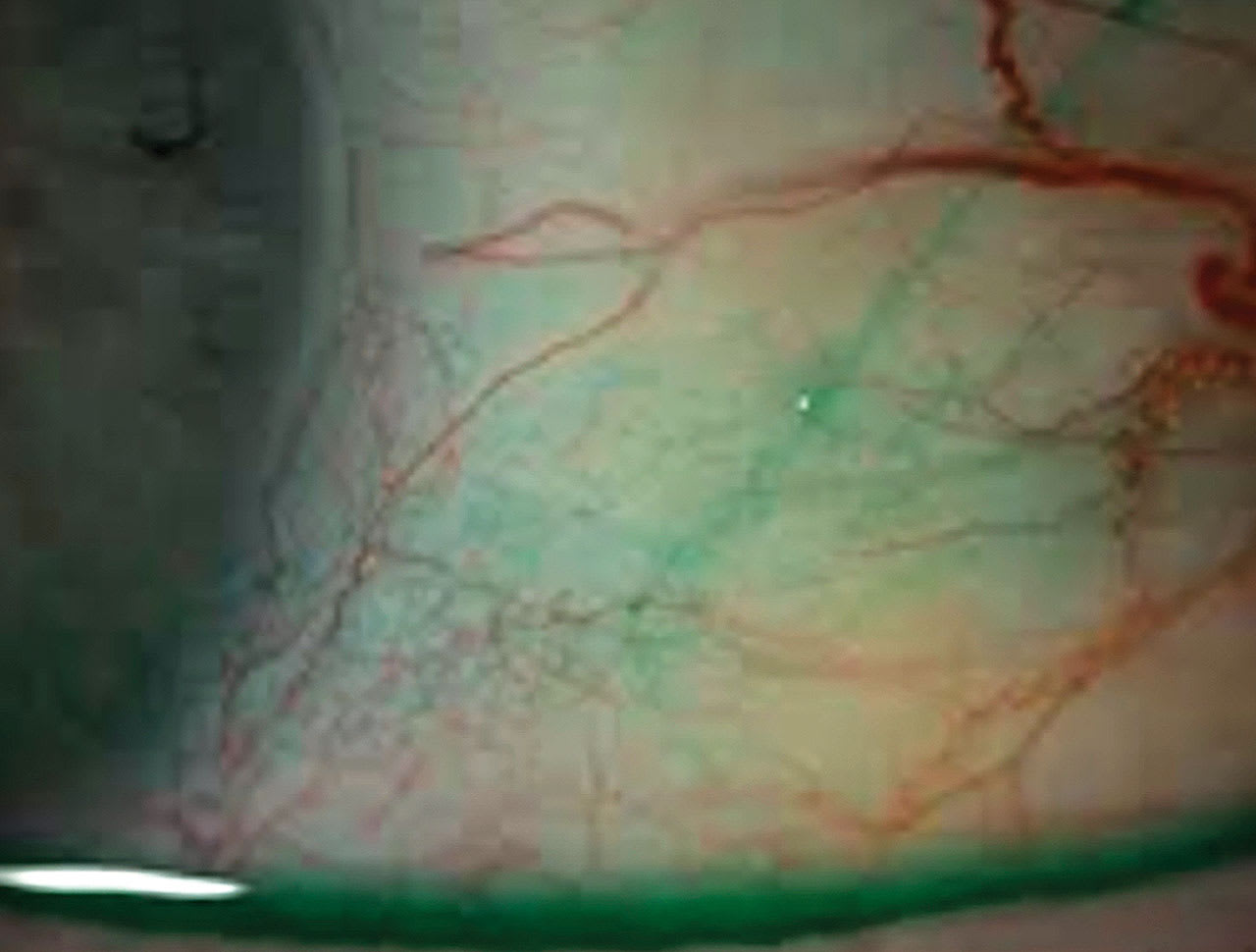
Lissamine green (LG) is a green dye that aids in the identification of DED and lid wiper epitheliopathy. Its primary ocular surface staining area is the conjunctiva. Slit-lamp observation using LG should be conducted with white light, 1 to 4 minutes after instillation. Staining visualization can be enhanced with either a Wratten 92 or a Hoya 25A filter. LG stains damaged or dead conjunctival cells and Marx’s line, including mucous strands. It also stains areas of disrupted intercellular junctions.
Personally, I find LG particularly useful for staging DED in patients whose symptoms do not have corneal fluorescein staining.
Combining Forces
A combination of 2% NaFl and 1% LG results in optimal staining without irritation.1 These combined dyes are available in both liquid and strip forms.
Reference
- Korb DR, Herman JP, Finnemore VM, Exford JM, Blackie CA. An evaluation of the efficacy of fluorescein, rose bengal, lissamine green, and a new dye mixture for ocular surface staining. Eye Contact Lens. 2008;34(1):61-64. doi: 10.1097/ICL.0b013e31811ead93.
