

Within the next few years, there will be an explosion of new presbyopia treatment options, such as intraocular lenses, multifocal contact lenses, and pharmaceutical agents. With this expansion of options, I anticipate an influx of presbyopic patients interested in trying them. Because the presbyopic population is at an increased risk for dry eye disease (DED) by virtue of their age, among other causes, this could be a golden opportunity for us to diagnose and treat DED in this population. Let’s dive in.
Shifting the Conversation
For some presbyopic patients, the onset of this refractive condition is the first time they are seeing an optometrist. Specifically, they may present to their OD with a complaint of unstable or fluctuating vision. This complaint can lend itself to shifting the conversation to DED; specifically, how a healthy ocular surface is required for optimal visual acuity. Regardless of the presbyopia treatment option the patient chooses, a robust tear film and healthy ocu-lar surface is necessary for clarity.
Some details to consider: An unhealthy ocular surface can negatively affect biometry readings for intraocular lenses. Multifocal contact lenses, as is the case with all contact lenses, can induce or exacerbate DED due to disruption to the natural tear film, among other causes. Additionally, the topical pharma-ceutical agents for presbyopia can contain ingredients that can influence ocular surface health.1
Incorporating Basic Diagnostics
After this conversation shift, we can incorporate basic diagnostic testing into the patient’s appointment to determine the possible presence of DED. A DED questionnaire is an efficient tool for seeing whether further diagnostic testing should be pursued. Various questionnaires are available. Some of the popular ones are the Dry Eye Questionnaire-5, Impact of Dry Eye in Everyday Life, Ocular Surface Disease Index (OSDI)-6, Standardized Patient Evaluation of Eye Dryness, and the Symptom Assessment in Dry Eye questionnaire. In the Tear Film & Ocular Surface Society Dry Eye Workshop III, the recommended questionnaire is the OSDI-6.2
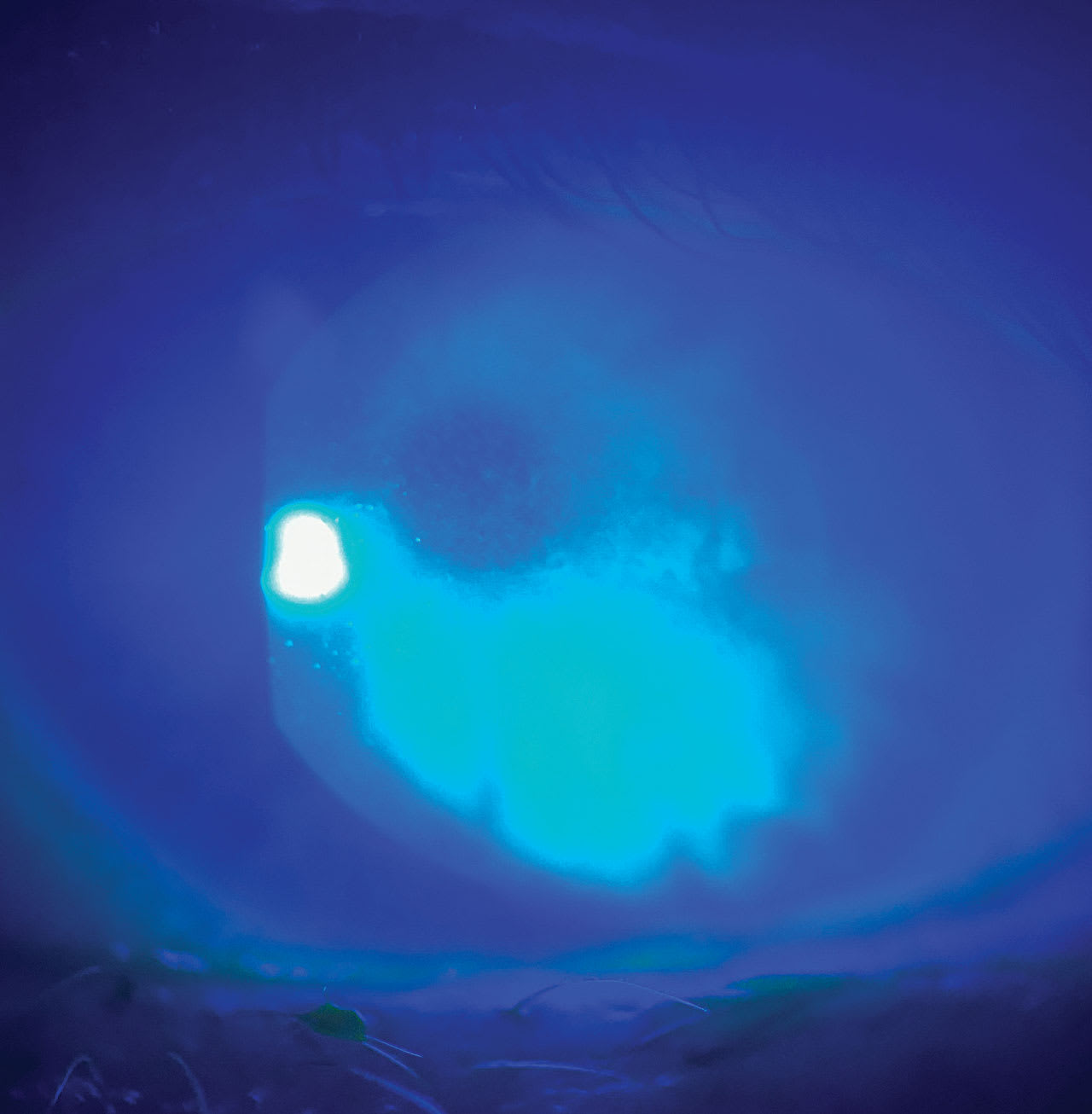
Further basic diagnostic testing includes corneal staining, tear break-up time assessment, Schirmer’s test, and manual meibomian gland assessment. From there, modern diagnostic tests can be used for diagnostic confirmation at a separate appointment if needed.
Other Discussions
I am hopeful that an increase in the awareness of presbyopia will also increase the frequency of eye exams in this working-age population of patients. Using this appointment as a discussion of presbyopia could also lend itself to conversations regarding the long-term effects of computer and digital device use on the meibomian glands and tear film. This could effortlessly lead to the opportunity to dive deeply into evaluating their ocular surface.
References
1. Zhang Y, Kam WR, Liu Y, Chen X, Sullivan DA. Influence of pilocarpine and timolol on human meibomian gland epithelial cells. Cornea. 2017;36(6):719-724. doi:10.1097/ICO.0000000000001181.
2. Wolffsohn JS, Benítez-Del-Castillo J, Loya-Garcia D, et al. TFOS DEWS III diagnostic methodology. Am J Ophthalmol. Published online May 30, 2025. doi:10.1016/j.ajo.2025.05.033doi:10.1016/j.ajo.2025.05.033.