


A 71-year-old male presented with sudden onset of a paracentral scotoma in the left eye while traveling abroad. He described a “blocked-out” region affecting the inferior central visual field that was more noticeable in dim lighting. Medical history was notable for well-controlled type 2 diabetes (A1C ~6.1) and historically low blood pressure (~104/70). One to two weeks prior to symptom onset, the patient reported several days of unilateral temporal headaches.
Case Study
Initial visual acuity measured 20/20 OD and 20/30 OS. Dilated fundus examination was largely unremarkable, revealing no retinal hemorrhage, edema, or vascular attenuation. A prior posterior vitreous detachment with Weiss ring was noted in the left eye. Visual field testing demonstrated a superior hemifield defect OS, though the patient subjectively reported an inferonasal scotoma.
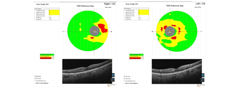
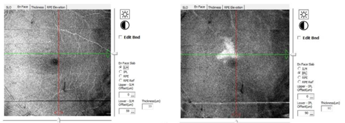
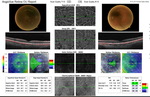
GCC thickness maps of the right and left eye demonstrated localized parafoveal changes corresponding to the ischemic area in the left eye (Figure 1). Optical coherence tomography (OCT) en-face imaging revealed a focal hyperreflective band within the inner retinal layers superior nasal to the fovea, localized to the inner plexiform layer (IPL) (Figure 2). These findings were consistent with Paracentral Acute Middle Maculopathy (PAMM), a form of retinal ischemia affecting the intermediate and deep capillary plexuses. Laboratory testing including erythrocyte sedimentation rate (ESR) and C-reactive protein (CRP) was within normal limits, reducing the likelihood of giant cell arteritis.
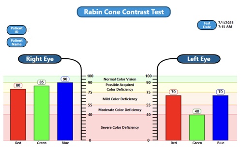
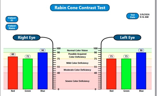
Initial cone contrast testing demonstrated reduced cone contrast sensitivity OS, particularly involving the green cone pathway, suggesting localized retinal dysfunction corresponding to the ischemic region (Figure 3).
The patient was counseled that the scotoma likely represented permanent retinal injury, though central visual acuity was expected to remain stable. Management focused on monitoring retinal structure and emphasizing control of systemic vascular risk factors.
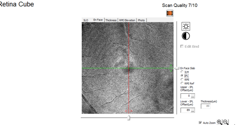
At follow-up several months later, visual acuity had improved to 20/20 OU, though the patient continued to perceive the paracentral scotoma. Repeat OCT imaging demonstrated resolution of the previously observed hyperreflective lesion within the IPL representing the transition from the acute ischemic phase of PAMM to the chronic stage following retinal infarction (Figure 4),
OCT angiography (OCTA) revealed persistent vascular abnormalities in the left eye compared with the right, including reduced superficial capillary vessel density and an enlarged foveal avascular zone (FAZ) (Figure 5). FAZ area measured 0.25 mm² OS compared with 0.15 mm² OD, confirming underlying retinal microvascular compromise.
Repeat cone contrast testing demonstrated substantial functional improvement, with cone contrast scores returning to levels comparable to the fellow eye (Figure 6). This suggests functional recovery within surrounding retinal tissue despite permanent damage to the localized infarct region. Electroretinography confirmed globally intact retinal function, supporting the conclusion that the visual deficit represents localized retinal injury rather than generalized retinal dysfunction.
Conclusion
This case highlights several important clinical features of PAMM. Fundus examination may appear normal despite significant visual symptoms, emphasizing the importance of OCT imaging in patients presenting with unexplained paracentral vision loss. While structural OCT findings often resolve over time, OCTA may reveal persistent vascular abnormalities corresponding to the original ischemic insult. Functional testing, such as cone contrast sensitivity, may demonstrate recovery in surrounding retinal tissue despite persistent subjective scotoma.
Multimodal imaging—including OCT, OCTA, and functional testing—provides valuable insight into the structural, vascular, and functional consequences of retinal ischemia.
Key Clinical Takeaways
A few key clinical takeaways from this case include:
· PAMM may present with normal fundus examination findings.
· OCT is essential for identifying inner retinal ischemia.
· OCTA can reveal persistent microvascular compromise after structural recovery.
· Functional testing may show partial recovery despite permanent scotoma.

This content is sponsored by Visionix